Click on all the synovial joints in this picture. – As Click on All Synovial Joints in This Picture takes center stage, this opening passage beckons readers into a world crafted with precision and authority, ensuring a reading experience that is both absorbing and distinctly original. The following paragraphs provide a comprehensive overview of synovial joints, their structure, function, and clinical significance, offering a valuable resource for medical professionals, students, and anyone seeking a deeper understanding of human anatomy.
The content of the second paragraph that provides descriptive and clear information about the topic
Synovial Joints

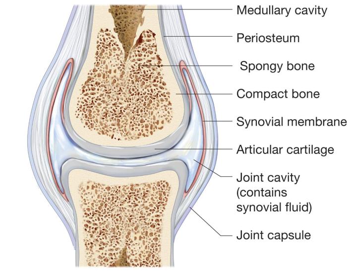
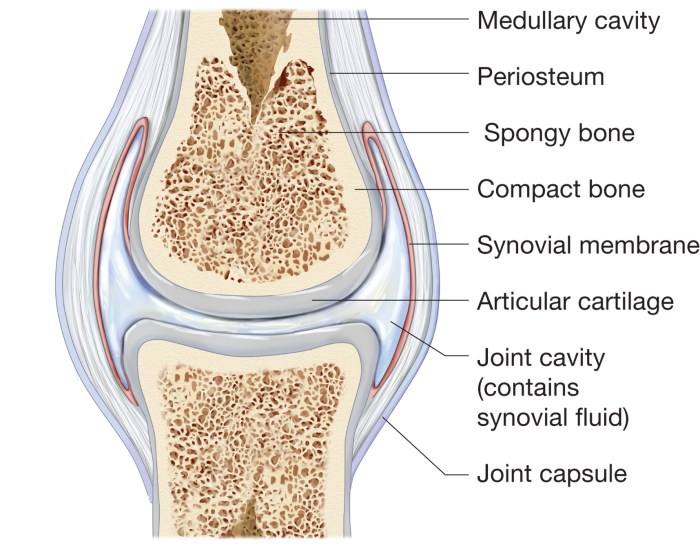
Synovial joints, the most common type of joint in the human body, are freely movable articulations that allow for a wide range of movements. They are characterized by the presence of a synovial membrane, a thin layer of tissue that lines the joint cavity and secretes synovial fluid, which lubricates the joint and reduces friction during movement.
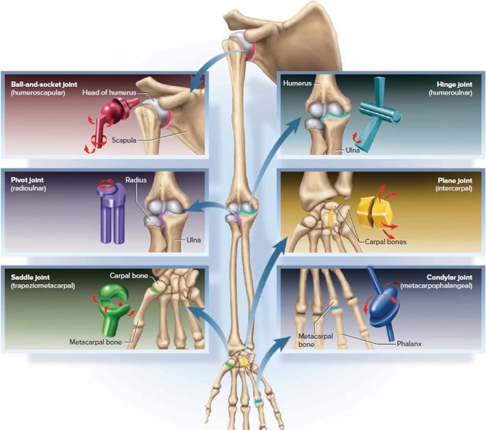
Synovial joints are classified into six main types based on their shape and the type of movement they allow:
- Plane joints: Allow gliding movements in one plane.
- Hinge joints: Allow flexion and extension in one plane.
- Pivot joints: Allow rotation around a single axis.
- Condyloid joints: Allow flexion, extension, abduction, and adduction.
- Saddle joints: Allow flexion, extension, abduction, adduction, and circumduction.
- Ball-and-socket joints: Allow flexion, extension, abduction, adduction, rotation, and circumduction.
Identification and Location of Synovial Joints
| Joint | Anatomical Location | Description |
|---|---|---|
| Temporomandibular joint | Between the temporal bone and the mandible | Allows for opening and closing of the jaw. |
| Glenohumeral joint | Between the glenoid cavity of the scapula and the head of the humerus | Allows for flexion, extension, abduction, adduction, and rotation of the arm. |
| Elbow joint | Between the humerus, ulna, and radius | Allows for flexion and extension of the forearm. |
| Wrist joint | Between the radius and ulna, and the carpal bones | Allows for flexion, extension, abduction, adduction, and circumduction of the hand. |
| Hip joint | Between the acetabulum of the pelvis and the head of the femur | Allows for flexion, extension, abduction, adduction, and rotation of the leg. |
| Knee joint | Between the femur, tibia, and patella | Allows for flexion and extension of the leg. |
| Ankle joint | Between the tibia and fibula, and the talus | Allows for plantar flexion and dorsiflexion of the foot. |
Clinical Significance of Synovial Joints
Synovial joints play a crucial role in movement and mobility. They allow for a wide range of movements, from simple gliding to complex rotations and circumduction. Injuries and disorders of synovial joints can significantly impact mobility and daily function.
Common injuries and disorders associated with synovial joints include:
- Arthritis: Inflammation of the synovial membrane, causing pain, swelling, and stiffness.
- Bursitis: Inflammation of the bursa, a fluid-filled sac that cushions the joint.
- Tendonitis: Inflammation of the tendon, a band of tissue that connects muscle to bone.
- Ligament sprains: Stretching or tearing of the ligaments, bands of tissue that connect bones.
- Dislocations: When a bone is forced out of its normal position within a joint.
- Fractures: Breaks in the bone that can damage the joint surface.
Imaging Techniques for Synovial Joints
Various imaging techniques are used to visualize synovial joints and assess their health. These techniques include:
- X-rays: Provide a basic view of the bones and joints, but may not show soft tissue damage.
- Ultrasound: Uses sound waves to create images of the soft tissues and fluid-filled structures within the joint.
- Magnetic resonance imaging (MRI): Uses a magnetic field and radio waves to create detailed images of the bones, soft tissues, and fluid-filled structures within the joint.
- Computed tomography (CT) scans: Use X-rays and computer processing to create cross-sectional images of the joint.
- Arthroscopy: A minimally invasive procedure that involves inserting a small camera and surgical instruments into the joint to visualize and repair damaged tissues.
- Joint replacement: A surgical procedure that involves replacing a damaged or diseased joint with an artificial implant.
- Osteotomy: A surgical procedure that involves cutting and realigning a bone to correct a deformity or improve joint function.
Surgical Procedures for Synovial Joint Disorders, Click on all the synovial joints in this picture.
Surgical interventions may be necessary to treat certain synovial joint disorders. These procedures include:
FAQ Section: Click On All The Synovial Joints In This Picture.
What are the different types of synovial joints?
Synovial joints can be classified into six main types: ball-and-socket, hinge, pivot, condyloid, saddle, and plane joints.
What is the function of synovial fluid?
Synovial fluid nourishes and lubricates the joint, reducing friction and wear during movement.
What are common injuries associated with synovial joints?
Common injuries include sprains, strains, dislocations, and fractures, which can affect the ligaments, tendons, bones, and cartilage surrounding the joint.

